
Medical 3d printing
Two-dimensional CT images to 3D printed parts
In cardiac surgery, CT, MRI, B-ultrasound and other examinations can only provide two-dimensional images. Doctors cannot see the whole picture of the heart directly, and cannot directly observe its internal fine structure. Therefore, it is difficult to grasp accurate and comprehensive patient information. Saina's WJP direct-injection color multi-material 3D printing technology can convert the patient's two-dimensional image data into a full-color 3D printing model of one-to-one size before the operation, vividly presenting the heart structure, blood vessels and other internal structures It is in front of doctors and can provide more rich information that is difficult to display in traditional imaging examinations, thereby greatly simplifying and standardizing the above-mentioned complex procedures, making the operation more accurate and safe.

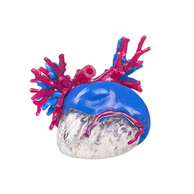
In cardiac surgery, CT, MRI, B-ultrasound and other examinations can only provide two-dimensional images. Doctors cannot see the whole picture of the heart directly, and cannot directly observe its internal fine structure. Therefore, it is difficult to grasp accurate and comprehensive patient information. Saina's WJP direct-injection color multi-material 3D printing technology can convert the patient's two-dimensional image data into a full-color 3D printing model of one-to-one size before the operation, vividly presenting the heart structure, blood vessels and other internal structures It is in front of doctors and can provide more rich information that is difficult to display in traditional imaging examinations, thereby greatly simplifying and standardizing the above-mentioned complex procedures, making the operation more accurate and safe.

The head is one of the most complex parts of the human body. Its environment is complicated and slender. In the reality that neurosurgery is difficult to implement, based on patient data obtained from medical examinations such as CT, with the help of Senna WJP direct-injection color multi-material 3D printing Technology, 3D printed a colorful, transparent, soft and hard model before surgery, clearly showing the anatomical position relationship between the lesion and the intracranial arteriovenous skull, facilitating the separation of intraoperative lesions and avoiding damage to intracranial blood vessels, thereby minimizing Surgery damage, improve the accuracy of the operation, help the smooth implementation of the operation.

The lungs are one of the most complex organs in the human body. According to medical examinations such as CT or PET-CT scans The patient data obtained from the investigation is used to print a color and transparent patient lung model with the help of the WJP direct-injection color multi-material 3D printing technology of Saina, so that the doctor can have an accurate understanding of the entire organ and lesion before the operation, and shorten the exploration time during the operation , Choose the most suitable surgical path, clearly determine where the lesion is in the lungs, the direction of blood vessels around the lesion, etc., and perform the operation more accurately.

In liver surgery cases, based on patient data obtained from medical examinations such as CT, the color and transparent liver model printed by the WJP direct-injection color multi-material 3D printing technology of Saina can accurately represent the overall shape of the liver, the lesion site, and clearly reproduce the liver interior The morphology of blood vessels and the shape of lesions help to simulate the surgical process before surgery, determine the approach of liver resection, the scope of resection, and calculate the remaining liver volume, and finally optimize the surgical plan.

Urology
In urology cases, color and transparent 3D printing models based on patient data obtained from medical examinations such as CT scans can be used to accurately and clearly mark different tissues in different colors, helping doctors accurately grasp the blood supply relationship between lesions and normal tissues. The doctor can simulate the surgical scene to the greatest extent before the operation, accurately evaluate the diseased condition, and plan the surgical path, so as to maximize the removal of the tumor while retaining the normal function of the organ, which can more effectively reduce the damage to normal tissues. lowest.

According to the patient’s CT or MRI scan data, use dedicated modeling software to complete the 3D model reconstruction, and print out the color, soft and hard combination of the patient’s bone lesions and injuries through the Senna WJP direct-injection color multi-material 3D printing technology. Print the model. Clearly and intuitively help doctors formulate surgical plans, complete surgical process planning, and assist in the design of personalized surgical instruments, which greatly reduces the occurrence of emergencies in the clinical operation stage and shortens the operation time.
Choose Your Printer
J 401
J 401 Pro
J 501 Pro
J 401
Supports printing of biocompatible materials
-
Support more than 10 million colors color scheme
-
Support 6 kinds of materials mixed printing at the same time
-
Resolution up to 14μm and 1800dpi printing accuracy
J401 Pro
Supports printing of biocompatible materials
-
Support more than 10 million colors color scheme
-
Support mixed printing of 7 materials at the same time
-
3 sets of high-performance print heads with 3840 nozzles for efficient printing
J501 Pro
Supports printing of biocompatible materials
-
Support more than 10 million colors color scheme
-
Support mixed printing of 7 materials at the same time
-
3 sets of high-performance print heads with 3840 nozzles for efficient printing

Bring Your Ideas to Life
Upload references or share your vision for a tailored technical quote. We help you transition from concept to finished print with professional precision.